Dr said my scan was normal" Answered by Dr Paxton Daniel Atelectasis Is lesswell inflated lung It can look like scarring orMay 27, 16 · The thorax is suited well to radiographic imaging due to the inherent subject contrast afforded by the airfilled lungs A vertical xray beam configuration should be used The craniocaudal area imaged should extend from cranial to the manubrium to at least one to two vertebral body lengths caudal to the most dorsocaudal limit of the diaphragmNotice the cranial location of the thoracic limbs relative to the thoracic inlet Figure 2 (A) Dog in left lateral recumbency with thoracic limbs pulled

Pyothorax Chez Un Chat Centre Hospitalier Clinique Veterinaire Cordeliers A Meaux 77
Radio thorax chat normal
Radio thorax chat normal-Thoracic radiographs of various dog breeds This section provides a web based overview of various normal dogs from a variety of dog breeds Almost all 71 dog breeds currently represented have at least 3 representative dogs of the same breed You can click on the individual images to zoom in from the case number home pageThorax thor´aks the part of the body between the neck and abdomen;


Radiographie Imagerie
Aug 02, 07 · The axial image demonstrates that the opacity on the chest film is actually the liver As we follow the livercontour, there is this unusual shape (yellow arrow) There is discontinuity of the crus which is a nonspecific sign (small blue arrow) On the axial image there is indentation of the liver on the posterior side due to blood in the thoraxCT chest (pre and post contrast, arterial phase) is the ideal investigation, to determine presence of aortic intramural haematoma, true lumen and extent of dissection Appearance There is a true lumen and a false lumen, separated by an intimal flapDec 01, 15 · Radiographic anatomy of the normal heart and the pulmonary vasculature The normal feline cardiac silhouette Thoracic radiography is one of the most commonly employed and useful tools in the diagnostic workup of cats with cardiac disease1, 2 It is used for differentiating cats with respiratory distress associated with cardiac disorders from those with respiratory
The thorax or chest is a part of the anatomy of humans, mammals, other tetrapod animals located between the neck and the abdomen In insects, crustaceans, and the extinct trilobites, the thorax is one of the three main divisions of the creature's body, each of which is in turn composed of multiple segments The human thorax includes the thoracic cavity and the thoracic wallApr 13, 21 · Thoracic wall The first step in understanding thorax anatomy is to find out its boundaries The thoracic, or chest wall, consists of a skeletal framework, fascia, muscles, and neurovasculature – all connected together to form a strong and protective yet flexible cage The thorax has two major openings the superior thoracic aperture found superiorly and the inferior thoracicThe normal thorax is well suited to radiographic evaluation because there is marked inherent contrast between the airfilled, fluidfilled, soft tissue, and bony structures that comprise the thoracic viscera and thoracic wall As has been stated before, at least 2 orthogonal views of the thorax are required for complete and accurate interpretation
"on a ct thorax what is dependent compressive atelectasis confirmed with prone and supine imaging?In women, they are 21 and 23 mm, respectivelyWhether thoracic radiographic findings can be used to aid clinicians in preliminarily differentiating the two tumor types before cytology or histopathology results become available Medical records, available cytologic or histologic samples, and thoracic


Clinique Veterinaire Des Bourgeolles Donzenac Brive Deroulement Radiographie Chien Chat Radiographie Veterinaire


Radiographies Du Chat
May 27, 16 · Principles of Radiographic Interpretation of the Thorax Donald E Thrall The acquisition of thoracic radiographs is common in small animal practice but is less common in the horse Computed tomography (CT) is very useful for characterizing intrathoracic disease in dogs and cats but is not used for thoracic imaging in horsesNormal CT C/ A/PA On the DorsoVentral view NO This is the DorsoVentral view In this case, the accessory lobe region is less aerated because of cranial displacement of the diaphragm secondary by the abdominal organs pressureThe apex of the heart is displaced on the left depending of the aspect of the thorax and the breed of the dog


Radiographies Du Chat
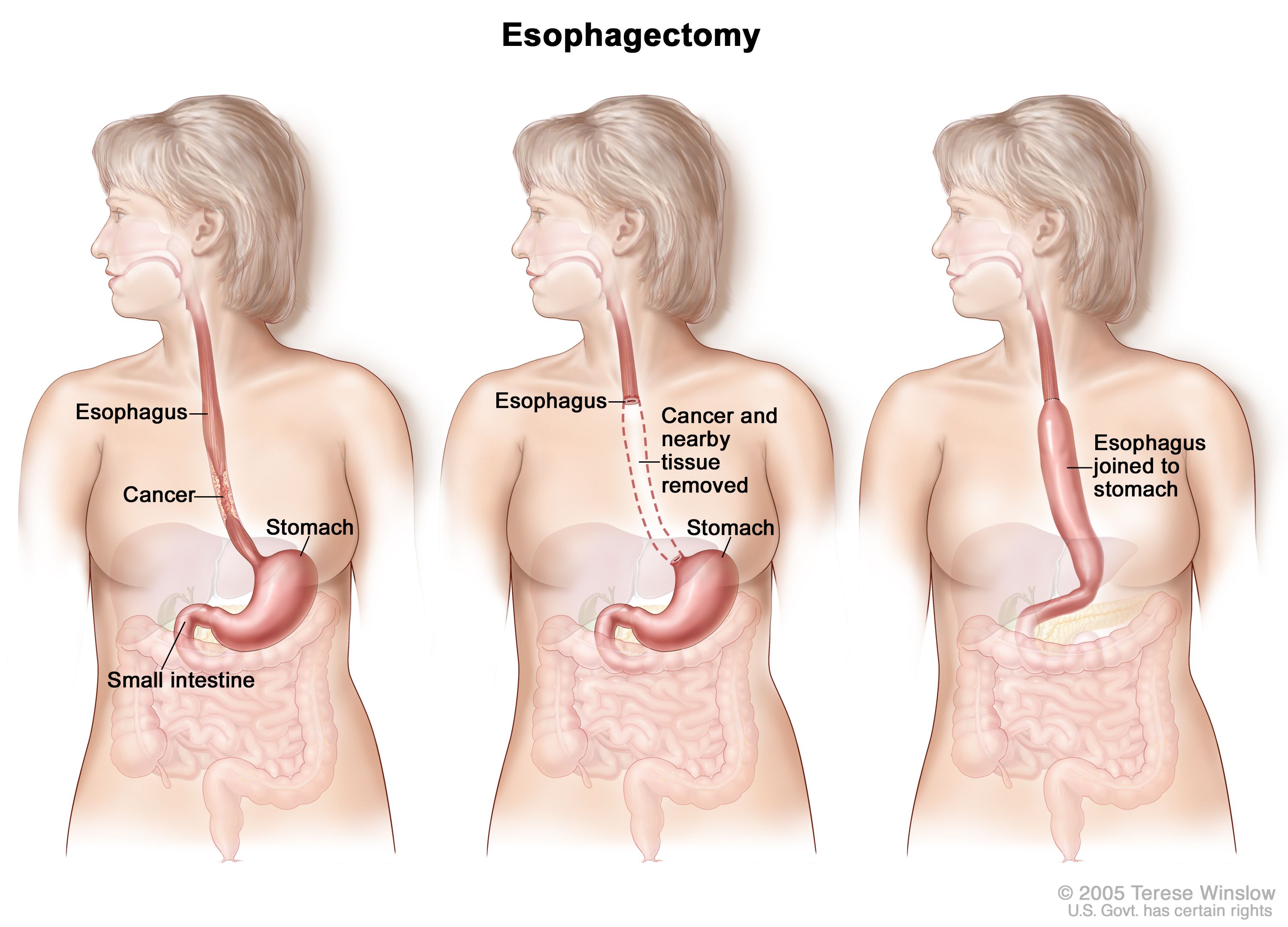


Esophageal Cancer Treatment Adult Pdq Patient Version National Cancer Institute
It is separated from the abdomen by the diaphragm Its walls are formed by the 12 pairs of ribs, attached to the sides of the spine and curving toward the front The principal organs in the thoracic cavity are the heart with its major blood vessels and the lungs with the bronchiNormal 2second CT anatomy of the thorax A (upper), Scan taken at hilar level Pulmonary vascular markings are shown throughout lung fields (white arrows) Descending branch of right pulmonary artery ( RP A) lies slightly lateral but predominantly anterior to air containing intermediate bronchusMay 06, 21 · Anatomy of the chest how to view the anatomical labels This atlas is a comprehensive and affordable learning tool for medical students and residents and especially for radiologists and pneumologists It provides access to CT images in the axial plane, allowing the user to learn and review the lung anatomy interactively


Radiographie Thoracique Images Normales Questions Et Images Fournises Par Dr Franck Durieux Dip Ecvdi Aquivet Radiographie Thoracique Images Normales Imagerie Diagnostique Et Outils Sessions Cardio Academy


111 Vertebral Heart Size Vhs Dr Buchanan S Cardiology Library Vin
UQ Med Yr 1 Chest Lungs and pleura;UNDER CONSTRUCTION This has two modules Learning Module To review the content of the subject Self assessment module You should use this module only after you have read textbooks or have knowledge of the subject and are ready to see whether you have comprehended the subjectUQ Med Yr 1 Introduction to Radiology;


Radiographie Veterinaire Centre Hospitalier Veterinaire Fregis


The Radiology Assistant Basic Interpretation
Nov 10, · Chest radiotherapy side effects Chest radiotherapy includes radiotherapy to the breast, your chest wall (if you've had surgery to remove your breast) or to your chest itself This can include radiotherapy to the lungs or to the oesophagus (your food pipe or gullet) Side effects will depend on where you're having treatment toUQ Radiology 'how to' series Chest CT Lungs and pleura;Thoracic Variant Anatomy War Machine;


Fractured Ribs Still Painful After 2 Months You May Need Surgery University Of Utah Health


Radiographie Thoracique Images Normales Questions Et Images Fournises Par Dr Franck Durieux Dip Ecvdi Aquivet Radiographie Thoracique Images Normales Imagerie Diagnostique Et Outils Sessions Cardio Academy
Canine Thorax Example 2 The following radiographs are the left lateral, right lateral and ventrodorsal views of the thorax of a tenyearold Mixed Breed Dog Metallic hemoclips are present in the cranial abdomenApr 02, 16 · The thymus is the dominant structure within the upper pediatric chest and is critical in the development of the immune system It is located in the anterior superior mediastinum and consists of two lobes that are fused in the midline The size, shape, and imaging finding of the normal thymus changes with ageThoracic radiography is one of the most commonly employed diagnostic tools for the clinical evaluation of cats with suspected heart disease and is the standard diagnostic method in the confirmation of cardiogenic pulmonary edema In the past, interpretation of feline radiographs focused on a descrip


Pyothorax Chez Un Chat Centre Hospitalier Clinique Veterinaire Cordeliers A Meaux 77


Radiographie Du Thorax Normale 1 Youtube
CT scan of Chest;A series of annotated radiographical images highlighting the key anatomical structures of the central nervous system head and neck spine thorax abdomen and pelvis upper limb lower limb Part of our Medical Imaging Anatomy Course OnlineAnswer CXR Outline mediastinum What is the normal width of mediastinum at supra cardiac vessel area?


Pneumothorax Et Pneumomediastins Spontanes Chez Un Chat Centre Hospitalier Clinique Veterinaire Cordeliers A Meaux 77


Pyothorax Chez Un Chat Centre Hospitalier Clinique Veterinaire Cordeliers A Meaux 77
The normal trachea at the thoracic inlet is between 15 and % of the thoracic inlet internal dimension as measured on the lateral radiograph For bulldogs and other brachycephalic breeds this measurement can approach 12% and still be considered normalJoshua Broder MD, FACEP, in Diagnostic Imaging for the Emergency Physician, 11 Cost of Chest Xray Chest xray is one of the most costeffective imaging examinations, with the cost to patients being between $50 and $2 in many medical systems 1 However, charges for chest xray vary considerably, even in the same geographic region According to data provided by health careThis CT scan of the upper chest (thorax) shows a malignant thyroid tumor (cancer) The dark area around the trachea (marked by the white Ushaped tip of the respiratory tube) is an area where normal tissue has been eroded and died (necrosis) as a result of tumor growth
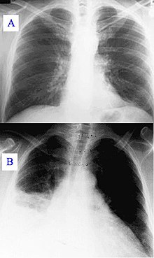


Radiographie Du Thorax Wikipedia


Radiographie Radioscopie Clinique Veterinaire Des Docteurs Martin Granel Beaufils Jumelle Calvisson
(Slideset Comment examiner une radio du thorax ?) slide 15 Lorsque plusieurs tubulures centrales se chevauchent, il peut être plus difficile de déterminer leur emplacement Ce cliché d'une radiographie thoracique portative AP montre une tubulure dans la veine jugulaire interne droite et une tubulure dans la veine sous clavière droite(A) Dog in right lateral recumbency with thoracic limbs pulled cranially See text for anatomic boundaries of collimated thorax (B) Right lateral thoracic radiograph of dog in Figure 2A;Radio du thorax Introduction Partie 1 Docteur SynapaseRéférences 1 https//radiopaediaorg2 http//wwwradiologyassistantnl
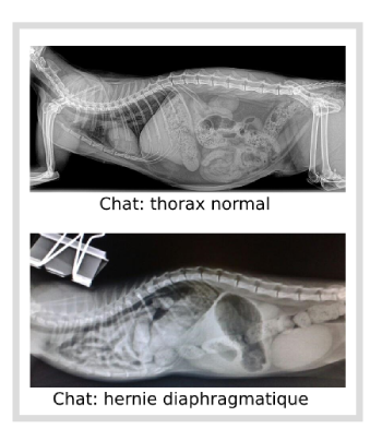


Blessures Chez Les Petits Animaux


Pyothorax Chez Un Chat Centre Hospitalier Clinique Veterinaire Cordeliers A Meaux 77
Feb 18, 13 · It is a normal finding, which can be seen on many chest xrays and should not be mistaken for pathology in the lingula or middle lobeJun 12, 15 · Figure Chest xray of a patient with pneumonia A, Posteroanterior view reveals a patchy opacity overlying the left lung base (large arrow)Note that the left cardiac border is preserved, indicating that the pneumonia is more likely in the left lower lobe If it were lingular and abutting the left cardiac border, the left cardiac border would be obscured (the "silhouette" sign)Lesions of the chest wall include trauma (fractures, swelling, SQ emphysema), infections, degeneration, and neoplasia Many chest wall lesions present as masses, and if they project into the thorax may demonstrate an extrapleural mass sign 1 Well defined convex border facing the pulmonary surface 2 Tapered edges which blend into the chest


Normal Lung X Ray Vs Copd Perokok C


Rayons X Des Jambes Du Thorax Et La Tete Avant D Un Chat Normal Image Stock Image Du Humerus Radiologie
Normal Radiographs If you are wondering whether what you're seeing is a lesion or not, check some normal radiographs for comparison Here is my list of normal cases for reference Thorax Normal Thorax 1 6 yearold, male neutered, canine Samoyed Normal Thorax 2 7 yearold, male neutered, canine WeimaranerAnswer CXR Identify Trachea, carina, right and left main stem bronchi Why are they visible, while rest of the bronchial tree is not?How to Read Chest Films • Develop a System – Doesn't matter what it is, just make sure you look at EVERYTHING • Look at a lot of films • Know the limits of the modality – Poor positioning, technique, motion, etc • Know some basic patterns What You Should Recognize • Normal •CHF • Consolidation • Effusions • Masses
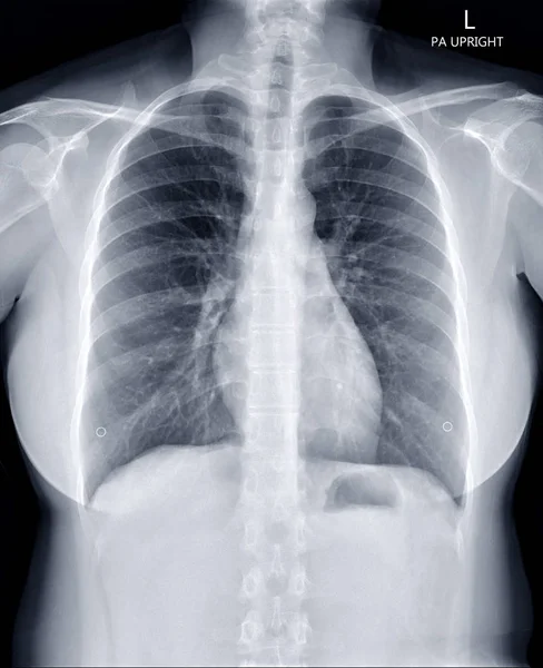


33 Cxr Stock Photos Images Download Cxr Pictures On Depositphotos


Les Tumeurs Chez Le Chat Chat Fait Du Bien
Feb 18, · This module of vetAnatomy is a basic atlas of normal imaging of anatomical feline radiology The 39 sampled xray images of healthy cats were performed by Susanne AEB Borofka (PhD dipl ECVDI, Utrecht, Netherland) Those images were categorized topographically into six chapters (head, vertebral column, thoracic limb, pelvic limb, thorax and4e année médecine – Rotation 3 – 15/16 ISM Copy Module de Cardiologie Interprétation d'une radiographie thoracique Télé thorax (TLT) de face se prend en inspiration forcée, les membres supérieurs en pronation forcée les paumes en dehors Télé thorax de profil se prend le coté malade sur la plaque, les bras levésWhat is the normal width of mediastinum at tracheal level?


Radiographies Du Chat


The Radiology Assistant Basic Interpretation
In summary in a normal chest Xray, anatomical borders of the peripheral bronchi are invisible However, due to pathologic changes bronchi can sometimes be distinguished When the alveoli are filled with fluid (blood, pus, mucus, edema, cells) rather than air, a density difference develops between the alveoli and bronchiJun 24, 19 · Thoracic radiography is an essential diagnostic technique for the diagnosis of intrathoracic and some systemic diseases It is also one of the most challenging areas of veterinary radiography, regarding radiographic technique and interpretation of the image so it is important to be familiar with the normal appearance of the structuresOn which view the accessory lobe region would be best observed ?
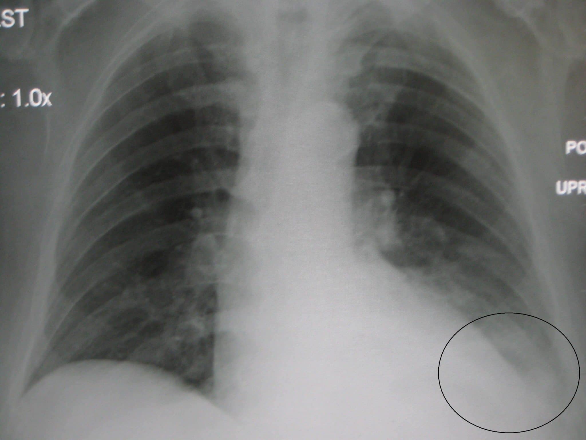


Chest X Ray Interpretation A Structured Approach Radiology Osce


Chat Radio
Sep 01, 01 · (a, b) Drawings (axial view) illustrate the normal thoracic CT anatomy at the level of the T2 (a) and T3 (b) vertebral bodies (c, d) Pancoast tumor in a 62yearold man with Horner syndrome (c) Axial CT scan demonstrates a Pancoast tumor in the left upper lobe (t) The mass is contiguous with the first rib in the area of the inferior cervicalJul 21, 19 · Calcification of the cartilage rings is a common normal finding in patients older than 40 years, particularly women, but it is seldom evident on radiographs (Fig 14) The upper limits of normal for coronal and sagittal diameters of the trachea in men are 25 and 27 mm, respectively;Normal pressure hydrocephalus, aqueductal stenosis, Chiari I malformation Brain MRI without contrast & CSF flow study (Acqueductal stroke volume measurement) Mass MRI without and with contrast MRI contraindicated CT without and with contrast Aneurysm or AVM "Screening" MRA Head (noncontrast) @ 3T CTA head with contrast


Normal Chest Ct Radiology Case Radiopaedia Org


Radiographie Radioscopie Clinique Veterinaire Des Docteurs Martin Granel Beaufils Jumelle Calvisson


The Radiology Assistant Basic Interpretation


Radiographie Imagerie


Radiographie Radioscopie Clinique Veterinaire Des Docteurs Martin Granel Beaufils Jumelle Calvisson


Radiographie Imagerie


Rayons X Des Jambes Du Thorax Et La Tete Avant D Un Chat Normal Image Stock Image Du Humerus Radiologie


Que Peut On Voir Ne Peut On Pas Voir A L Examen Radiographique Clinique Veterinaire Mairie D Issy


Malignant Pleural Effusion Pulmonology Advisor
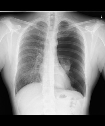


Pneumothorax Radiology Reference Article Radiopaedia Org


Radiographie Thoracique Images Normales Questions Et Images Fournises Par Dr Franck Durieux Dip Ecvdi Aquivet Radiographie Thoracique Images Normales Imagerie Diagnostique Et Outils Sessions Cardio Academy


Que Peut On Voir Ne Peut On Pas Voir A L Examen Radiographique Clinique Veterinaire Mairie D Issy


111 Vertebral Heart Size Vhs Dr Buchanan S Cardiology Library Vin
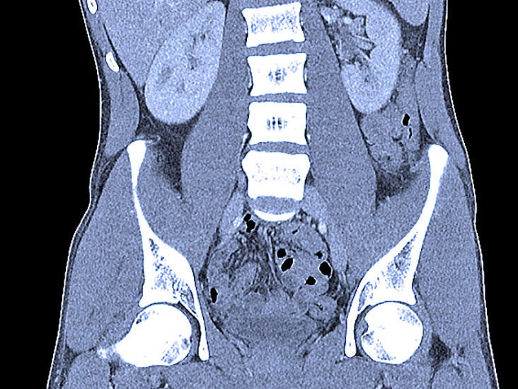


Abdominal Ct Scans Definition Uses Picture And More


You Don T Like Skydiving And I Don T Like Sex Why We Need To Talk About Asexuality The Correspondent


Radiographie Imagerie


Radiographie Radioscopie Clinique Veterinaire Des Docteurs Martin Granel Beaufils Jumelle Calvisson


Radiographie Boules De Fourrure
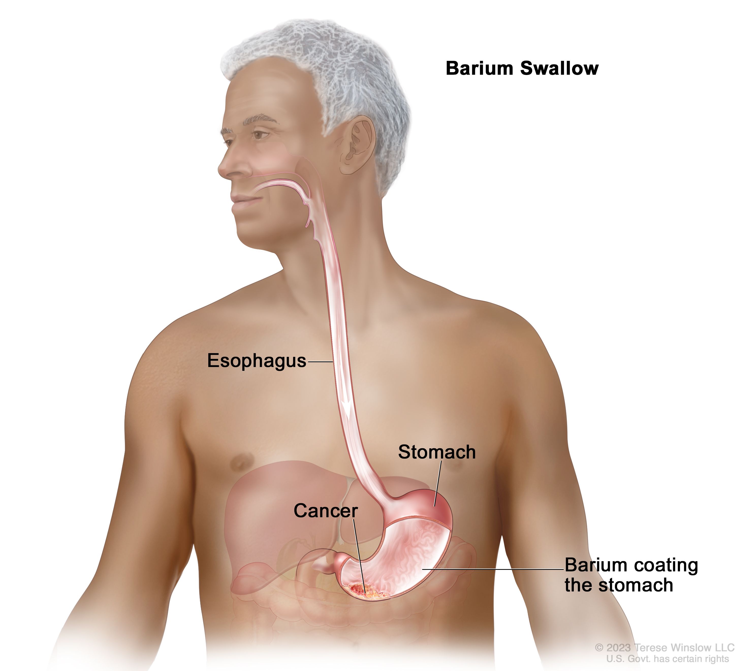


Gastric Cancer Treatment Pdq Patient Version National Cancer Institute


Pyothorax Chez Un Chat Centre Hospitalier Clinique Veterinaire Cordeliers A Meaux 77


Pdf Imaging Adults On Extracorporeal Membrane Oxygenation Ecmo


Neuroimaging Findings In Pediatric Genetic Skeletal Disorders A Review Wagner 17 Journal Of Neuroimaging Wiley Online Library


Normal Chest Imaging Examples Radiology Reference Article Radiopaedia Org


Auscultation Reconnaitre Le Bruit Bronchique Portail Video De L Universite De Rouen Normandie


Radiographies Du Chat


Radiographie Boules De Fourrure


œdeme Pulmonaire Cardiogenique Du Chat


Radiographie Radioscopie Clinique Veterinaire Des Docteurs Martin Granel Beaufils Jumelle Calvisson
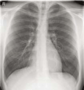


Thoraxfoto Hartwijzer Nvvc
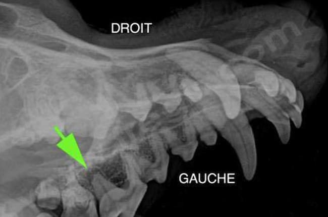


Radiographie Veterinaire Centre Hospitalier Veterinaire Fregis


Radiographies Du Chat


Radiographie Du Thorax Et De L Abdomen Du Chien Et Du Chat Le Point Veterinaire Fr


Radiographie Radioscopie Clinique Veterinaire Des Docteurs Martin Granel Beaufils Jumelle Calvisson


Radiographie Radioscopie Clinique Veterinaire Des Docteurs Martin Granel Beaufils Jumelle Calvisson


Radiographie Radioscopie Clinique Veterinaire Des Docteurs Martin Granel Beaufils Jumelle Calvisson


Radiographies Du Chat


Radiographie Boules De Fourrure


Radiographie Imagerie


Cours Pneumologie Aide A La Lecture D Une Radiographie De Thorax


Radiographie Radioscopie Clinique Veterinaire Des Docteurs Martin Granel Beaufils Jumelle Calvisson


Radiographie Boules De Fourrure


Thorax Radiologic Anatomy


Radiographie Radioscopie Clinique Veterinaire Des Docteurs Martin Granel Beaufils Jumelle Calvisson


Dyspnee Chez Un Chat


Cardiologie Clinique Veterinaire Des Docteurs Martin Granel Beaufils Jumelle Calvisson


Thorax Radiologic Anatomy


The Radiology Assistant Basic Interpretation


Que Peut On Voir Ne Peut On Pas Voir A L Examen Radiographique Clinique Veterinaire Mairie D Issy


Radiographie Radioscopie Clinique Veterinaire Des Docteurs Martin Granel Beaufils Jumelle Calvisson


Radiographie Thoracique Images Normales Questions Et Images Fournises Par Dr Franck Durieux Dip Ecvdi Aquivet Radiographie Thoracique Images Normales Imagerie Diagnostique Et Outils Sessions Cardio Academy


Radiographie Clinique Veterinaire Saint Francois


Clinique Veterinaire Des Bourgeolles Donzenac Brive Deroulement Radiographie Chien Chat Radiographie Veterinaire


Pdf Imaging Adults On Extracorporeal Membrane Oxygenation Ecmo
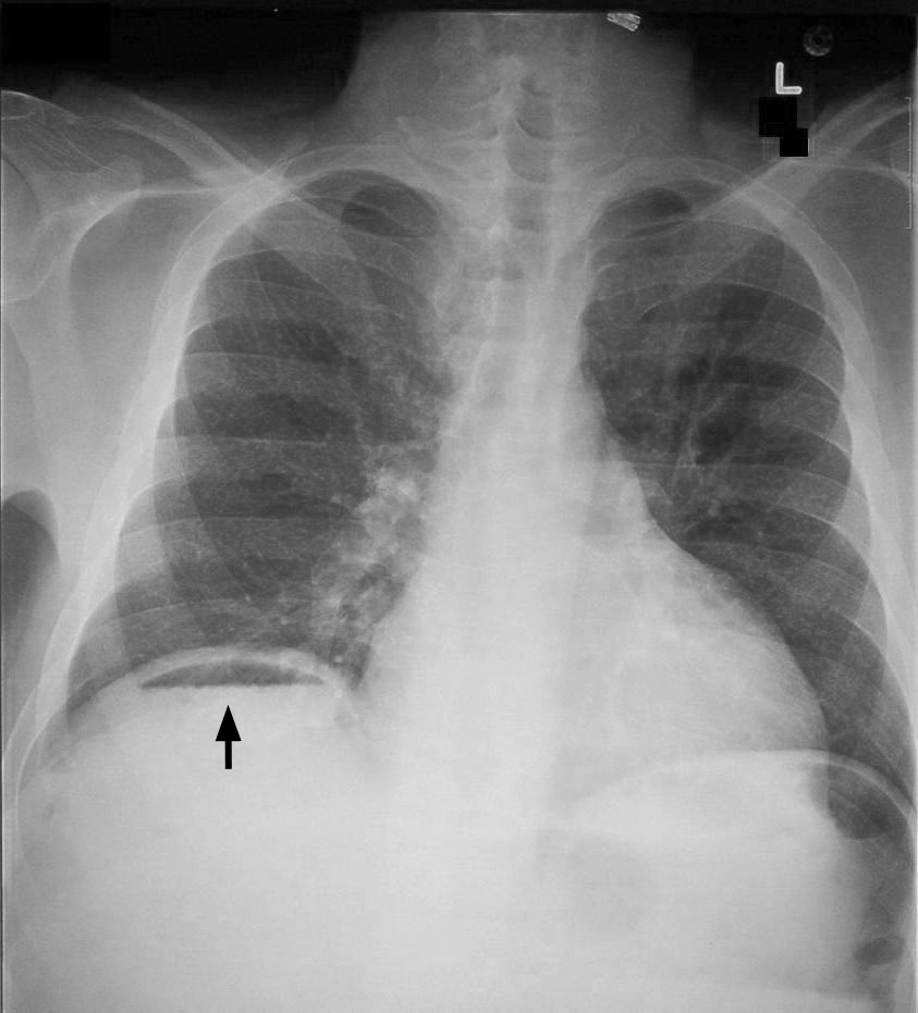


Chest X Ray Interpretation A Structured Approach Radiology Osce


Adenocarcinome Pulmonaire Chez Une Jeune Chatte Centre Hospitalier Clinique Veterinaire Cordeliers A Meaux 77


œdeme Pulmonaire Cardiogenique Du Chat


Cours Pneumologie Aide A La Lecture D Une Radiographie De Thorax
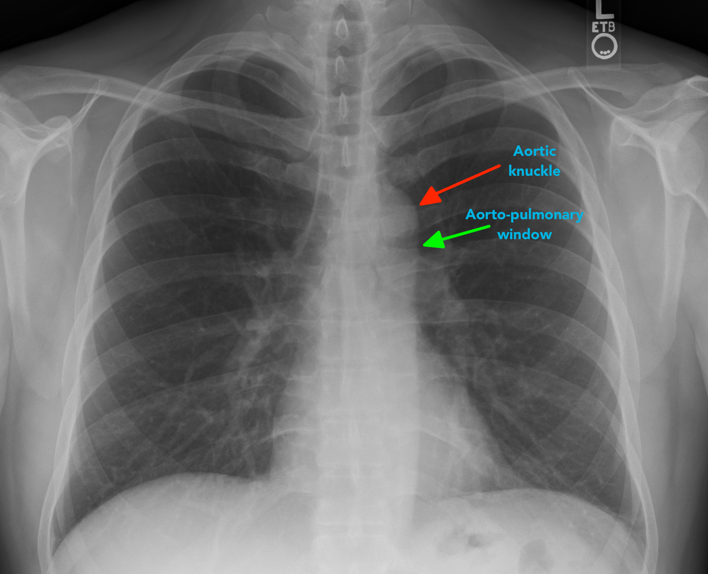


Chest X Ray Interpretation A Structured Approach Radiology Osce


Thorax Radiologic Anatomy


Radiographie Radioscopie Clinique Veterinaire Des Docteurs Martin Granel Beaufils Jumelle Calvisson
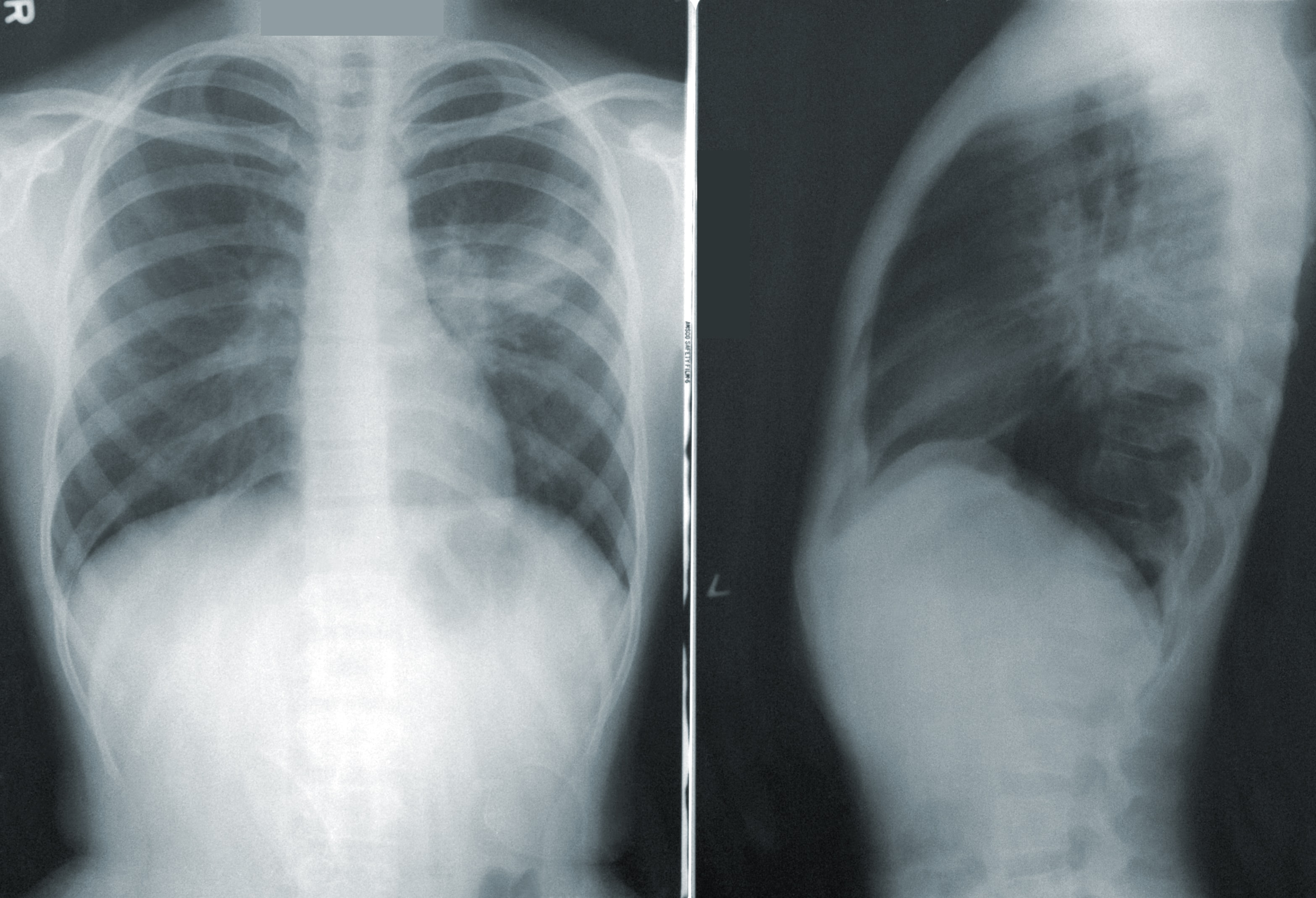


A Neural Network Can Help Spot Covid 19 In Chest X Rays Mit Technology Review


Dyspnee Chez Un Chat


Cours Pneumologie Aide A La Lecture D Une Radiographie De Thorax


Aucun commentaire:
Publier un commentaire